Methods: Wide subjects and new grounds
Mobile XRF analysis
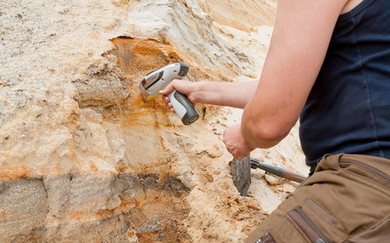
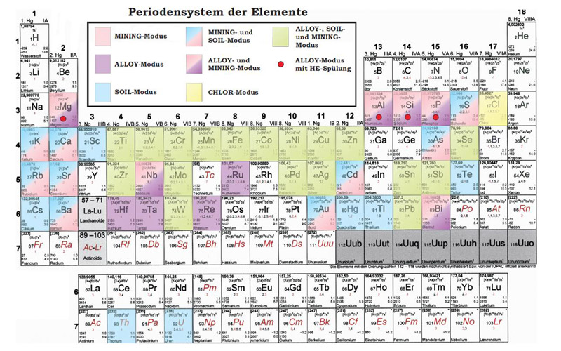
X-ray fluorescence analysis belongs to the modern standard operations in analytic chemistry. Since a few years this analytical method is utilized in the form of handheld devices usable in field studies.
Since 2008 we use equipment of the company Thermo Niton, more precisely the device XI3t. Crucial was the possibility to accurately assay sulfur in lignite starting from 0.1 mass%. Since then a regular mapping of the sulfur content takes place to display refined lignite in the seams.
An advantage of the XRF analysis is the possibility of simultaneous measurements of an entire set of elements. These range from aluminum to lead with contents of a few ppm up to decimal power values. Depending on the matrix of the measured sample different calibration modes exist, which allow various areas of application.
Applications of mobile XRF analysis
- Analysis of alloys
- Trace element analysis in soils and silica dominated samples (10 – 1000 ppm)
- Analysis of ores and concentrates (0.05 – 100 mass%)
- Analysis of elements in light, carbon-based samples (plastics, wood, coal, lignite)
- Possibility of personal calibration modes for any elements
Regarding the possibility of measuring almost unlimited numbers of points and as a consequence thereof the high representation, this method is a useful addition to the wet chemical analyses in applied field analytics.
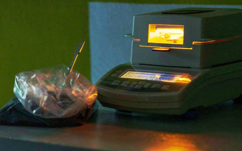
Further, the equipment can be used in the laboratory. Attributable to pretreatment of samples (drying, fine grinding) and a far longer measurement time the accuracy of the analytical method increases. The validation of accuracy happens by measuring certificated standards.
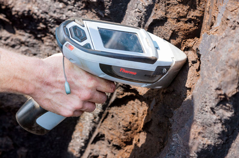
Due to the wide spectrum of elements and the coupling of fast analysis and field methods new fields of application arise. These range from analyses in the laboratory up to element screening on rock faces, trenches or drill cores. For further development of this promising method we rely on the creativity of our employees as well as our business partner.
To Geology »
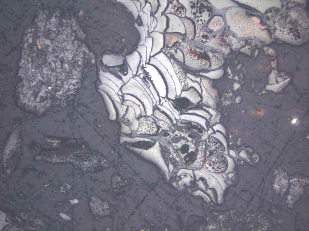
Microlithotype analysis (QMAT)
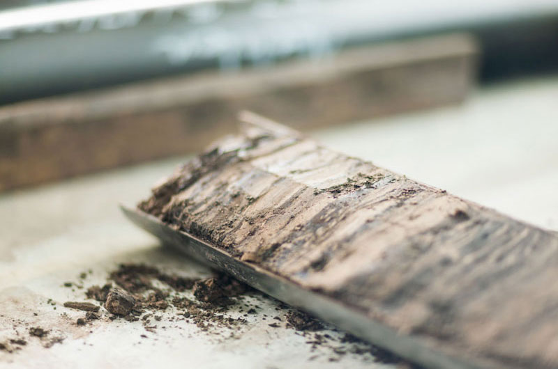
Quantitative microlithotype analysis (QMA) of soft lignite after the method of SONTAG/TZSCHOPPE/CHRISTPOPH (1965) was performed routinely since 1968 and upgraded in the year 1980 by SCHNEIDER to QMAT (quantitative microlithotype analysis with evaluation of textite).
Mechanical properties of lignite can be explained by microscopic recognizable structural features. Hence the suitability of various lignites for mechanical refining, like milling and agglomeration, is determinable by microscopic evaluation of structural features. The mechanical efficient microscopic structural area is defined as the microlithotype.

To Microscopy »
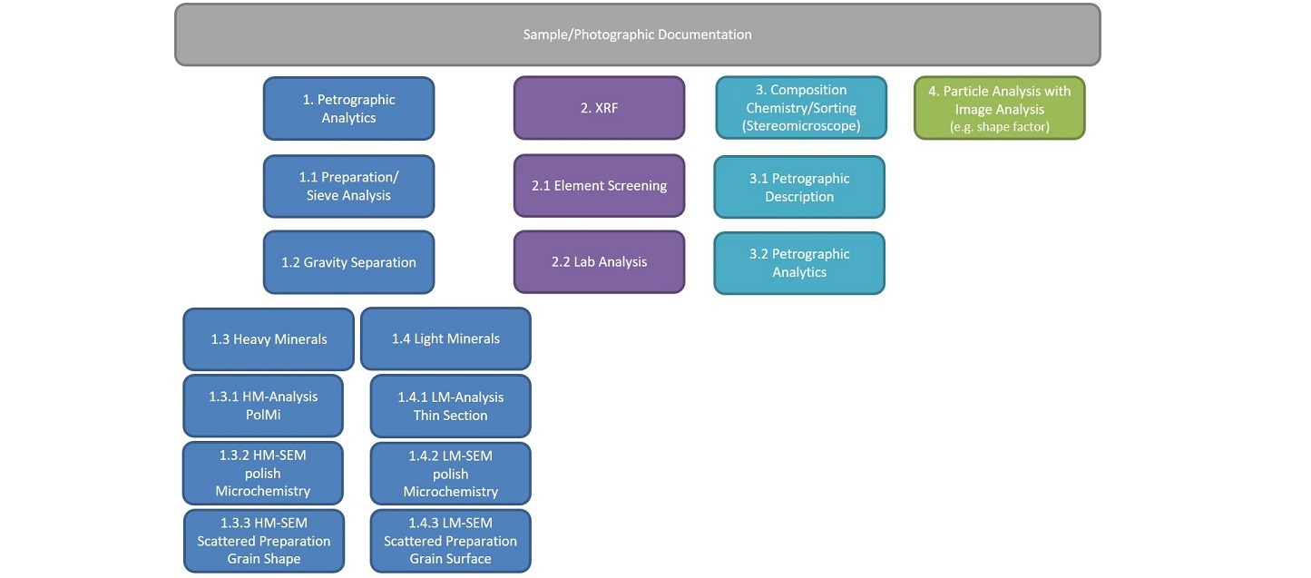
Particle analysis
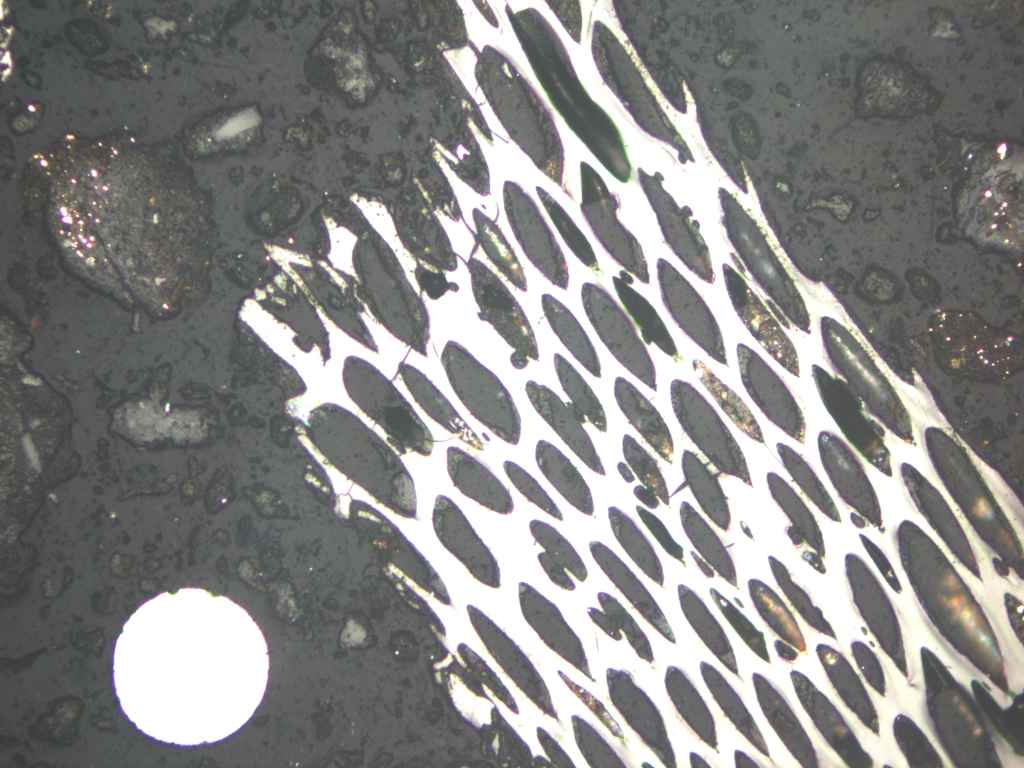
At particle analysis we deal with the spatial and material description of solids with a size of a few mm up to nanometer size. The range of tasks reaches from natural and artificial fibers to deposits in fluidly affected plants to analysis of dust in surroundings or at the workplace.
The advantage of particle analysis at LAOP is the combination of light optical, electron optical and microchemical methods, which make a profound sample description possible. Alternatively, the appropriated method for your problem can be chosen specifically from our analytical range.
Applications of Particle Analysis
- Morphological and microchemical characterization of dusts at industrial as well as settlement areas
- Analysis of deposition in pipelines
- Investigation of fibers from filter systems
- Description of tribological particles in hydrocarbon containing fluids (engines, generators, hydraulics)
- Analysis of fine sediments in natural and artificial waters
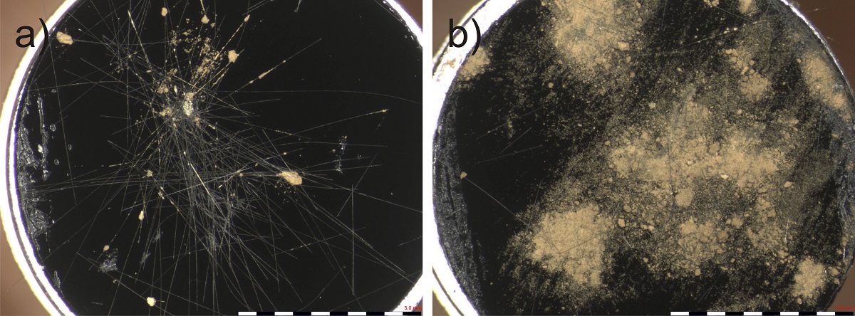
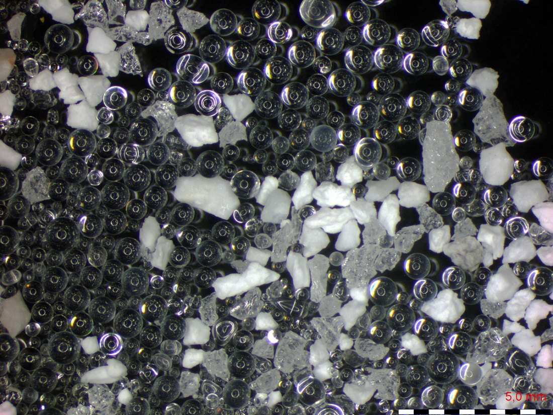
A particle analysis at LAOP can be realized qualitatively as well as quantitatively. Parameters in focus are: size, shape, surface and chemical composition.
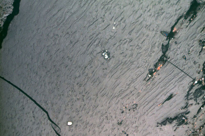
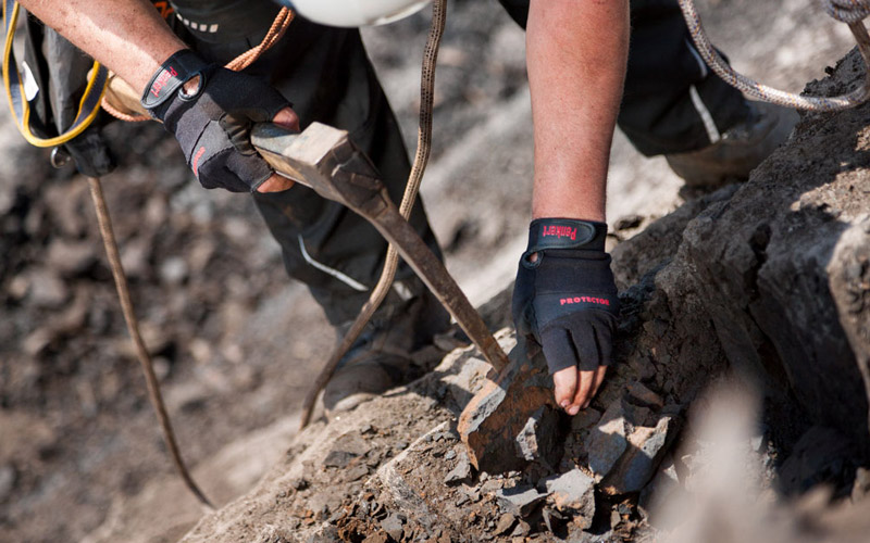
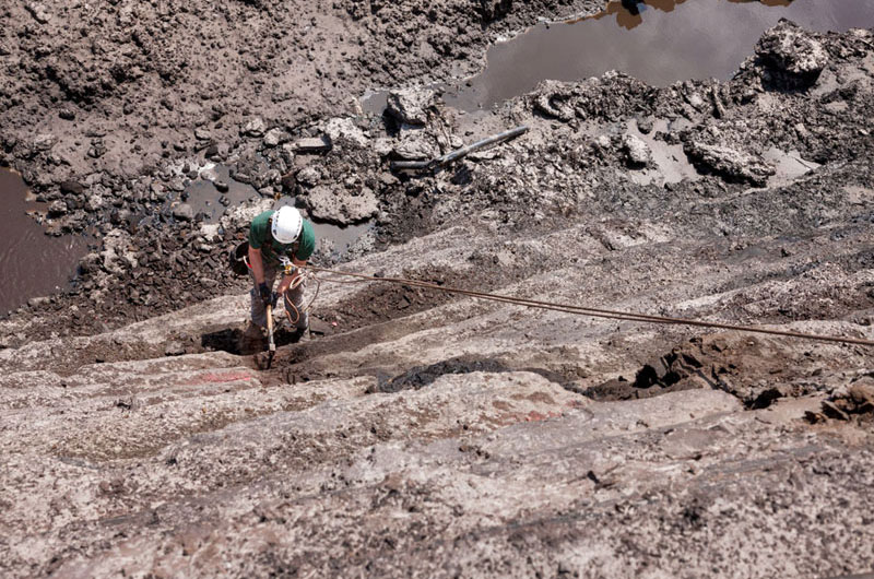
Rope-supported mapping

At the mapping in poorly accessible areas it needs special working methods for the purpose of health and safety aspiring minimal hazard potential. Those were developed by us with the help of rope-supported mapping. The rope-supported work primarily originates from the area of industry and plant construction.
It enables us to get access to seam parts were hoisting technology can't be applied. Further it is a cost-effective alternative to heavy technology.
Our employees which are familiar with this method are certified by FISAT as rope access technicians. All of our regular activities come to an agreement with the competent safety authority and will be incorporated in a job control statement. For here, too, is considered: SAVETY is a huge priority. With the method of rope-supported working we can offer all our services in the area of field geological mapping and sampling as well as field analytic by XRF even in exposed positions.

To Geology »
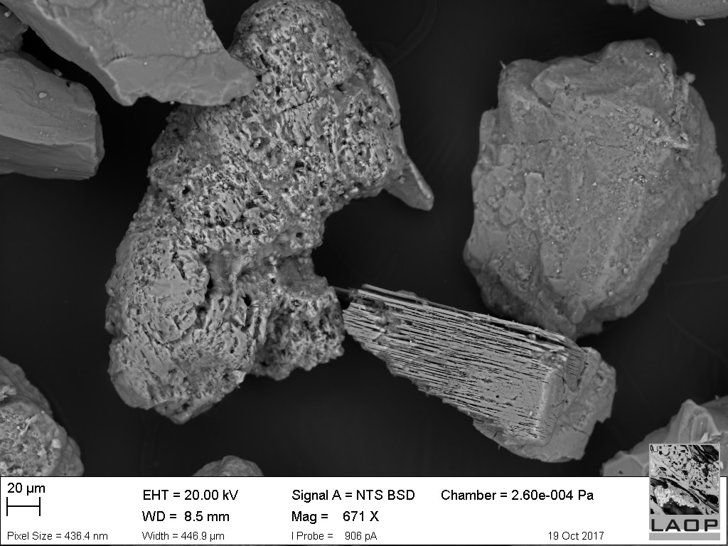
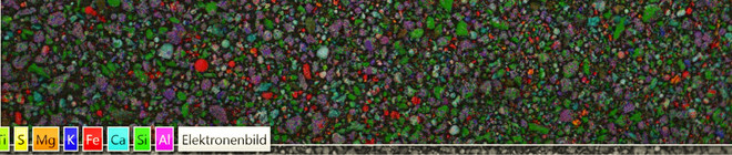
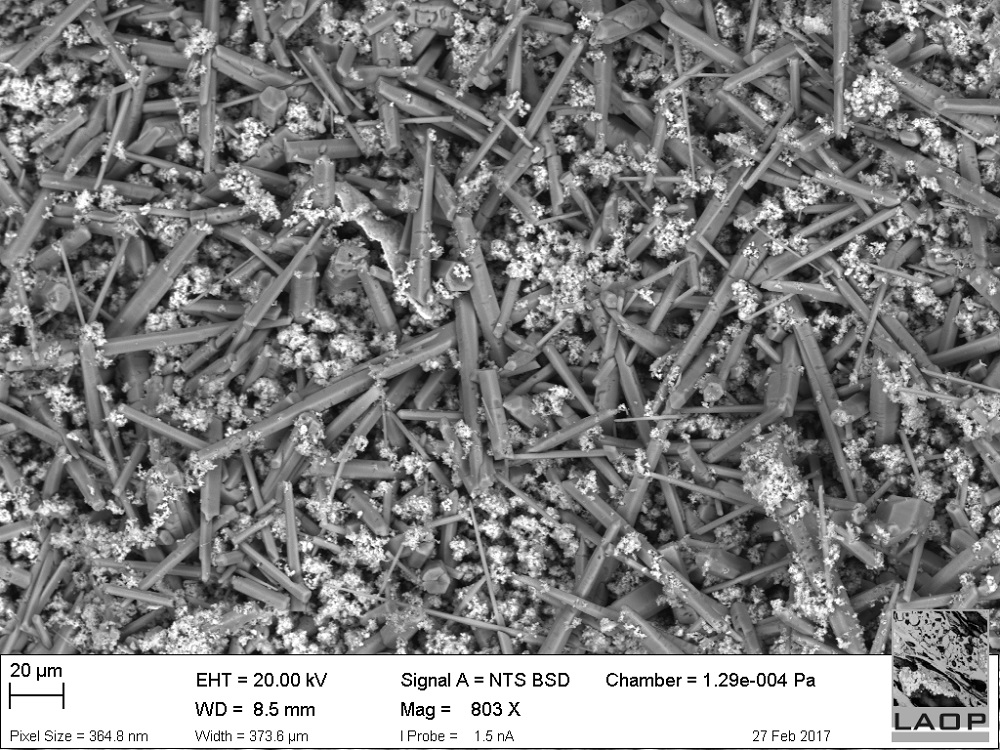
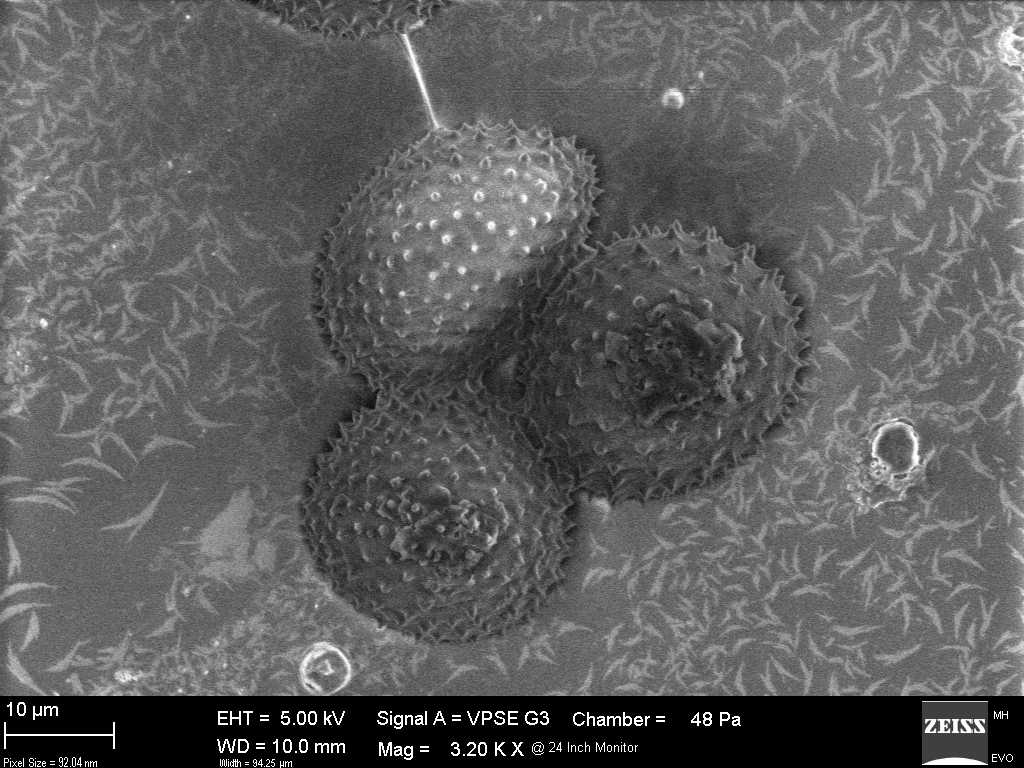
SEM
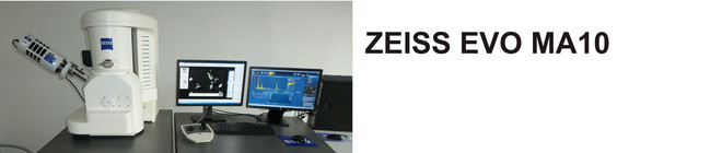
ScanningElectronMicroscopy is a profound method for the illustration and analysis of micro structures since more than 30 years. It supplies data about surface geometry, mineral content or crystallography of the sample material in a much higher resolution spectrum as light microscopy. Further a higher level of automation of analysis can be reached which leads to significant increase of accuracy and saving of time.
The technical progress and an economic production of micro electronic devices do not confine electron microscopy to university or research institution anymore. Today such equipment can be found in laboratories of industry, medicine and material science.
At LAOP investigation of palynology, heavy mineral analysis, paleobotany and micro chemistry are supported by the Zeiss EVO MA 10 with Oxford X-max 50 EDX.
Applications of ScanningElectronMicroscopy
- Illustration and comparison of recent and fossil spores and pollen, for example as basis of stratigraphic classification
- Micro chemical classification of the composition of heavy minerals as an aid for provenance analysis or for the prospection of placer deposits
- Analysis of slag, cinder and coverings of surfaces resulted from power plant processes
- Analysis of type, origin and micro chemistry of diverse dusts, for example caught particles from particle air drift experiments or fine particulate matter in inhabited areas
- Analysis and comparison of fossil and recent woods by means of wood-anatomic characteristics
- Image of all micro structures of geological, archeological and environmentally relevant samples

The installed SEM at LAOP works with a LaB6-cathode and reaches resolutions of 2 nm at 30 kV. It disposes a mode of operation with variable pressure for poorly conducting or degassing samples. For improvement of the samples conductivity and therefore for the taking of high-resolution images, coating techniques from Cressington are applied. Those are designed as dual-kit and enable a carbon coating as well as a coating with gold or platinum. Apart from the electron microscopic images, geometric measurements right up to volumetric analysis can be done. A real 3D surface picture can be achieved by means of the 5-segment-HD backscatter detector.
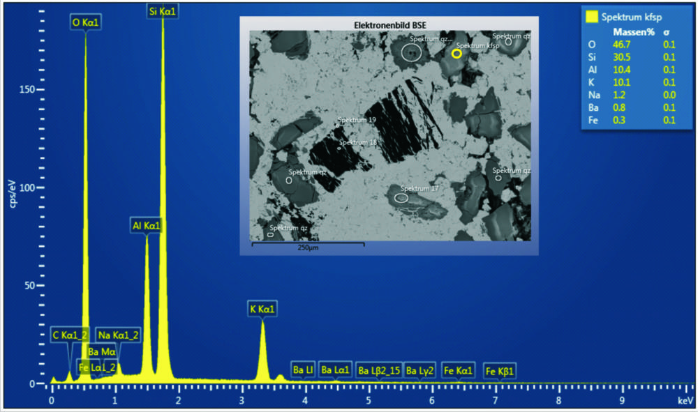
The EDX-detector enables point and line analysis as well as element mapping. This widens one’s understanding of the present sample material by the micro chemical composition and therefore complements our methods of light microscopy. Classification and presentation of individual phases as well as an automated particle analysis are optional features.
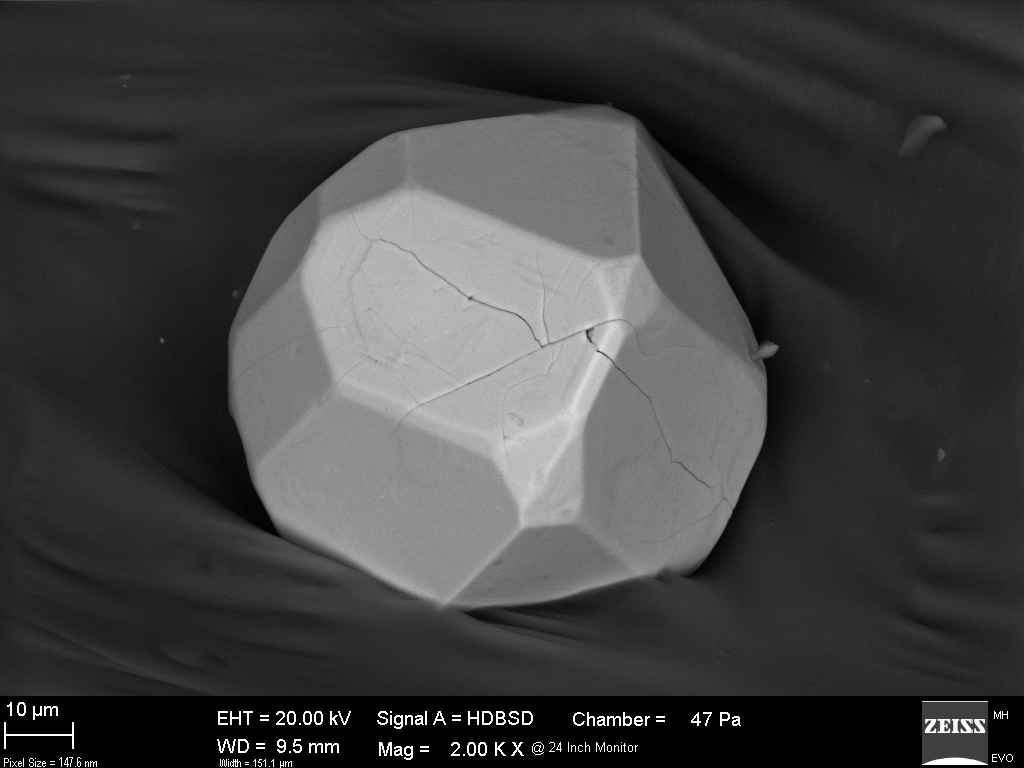
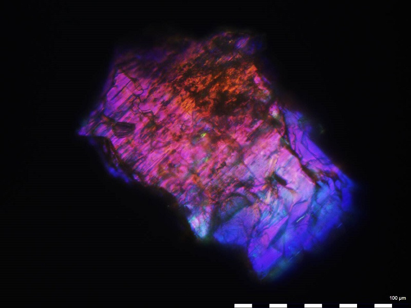
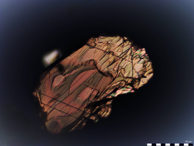
Heavy mineral analysis
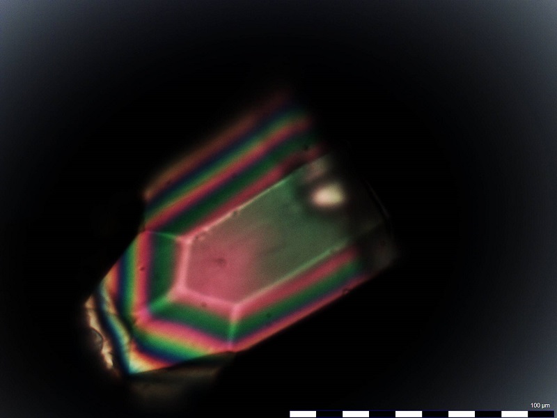
Since several years we intensively deal with the analysis of heavy minerals. In the process the sand is treated chemically and sieved subsequently. Now, for each obtained grain size range the light minerals are separated from the heavy ones. Latter, the group of heavy minerals, is characterized by a density of >2,9 g/cm³. The separation takes place in a separation funnel by use of the dense liquid sodium polytungstate. Opposite other dense liquids, sodium polytungstate has essential advantages. It is not toxic, not flammable, easy to handle and reusable.
Out of the separated sample a grain preparation is produced and microscoped subsequently. Within the group of heavy minerals transparent and opaque minerals can be distinguished. By use of transmitted-light microscopy only the former can be identified. The opaque minerals are only counted as a separate mineral group. For a more detailed identification we use a scanning electron microscope.
An heavy mineral fraction should have at least 300 grains, which will be mounted on an object slide. As mounting media we are using Meltmount (refractive index n=1,662). Counting and identification of the minerals on the polarization microscope takes place considering optical characteristics like color, cleavage, pleochroism, extinction, refraction and birefringence as well as conoscopic figure.
Applications of heavy mineral analysis
- Petrographic characterization of the sediments
- Make a point about stratigraphy, which can be indicated by mineral changes within a drill core or geological section
- Investigation of the paleogeographic development of sedimentation areas, including conclusions on delivery area, transport, tectonics, diagenesis and climate
- Determination of the content of rare earth elements, which are preferably enriched in heavy mineral placer
For the last application, apart from our SEM, our mobile XRF analysis is employed too. It is able to identify yttrium, lanthanum, cerum, neodymium and praseodymium from % up to the middle ppm range. Consequently the exploration of yttrium earth metals or cerite earth metals on drill cores or geological sections can be supported.

To Microscopy »
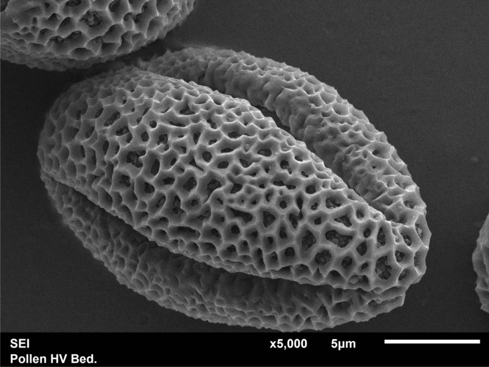
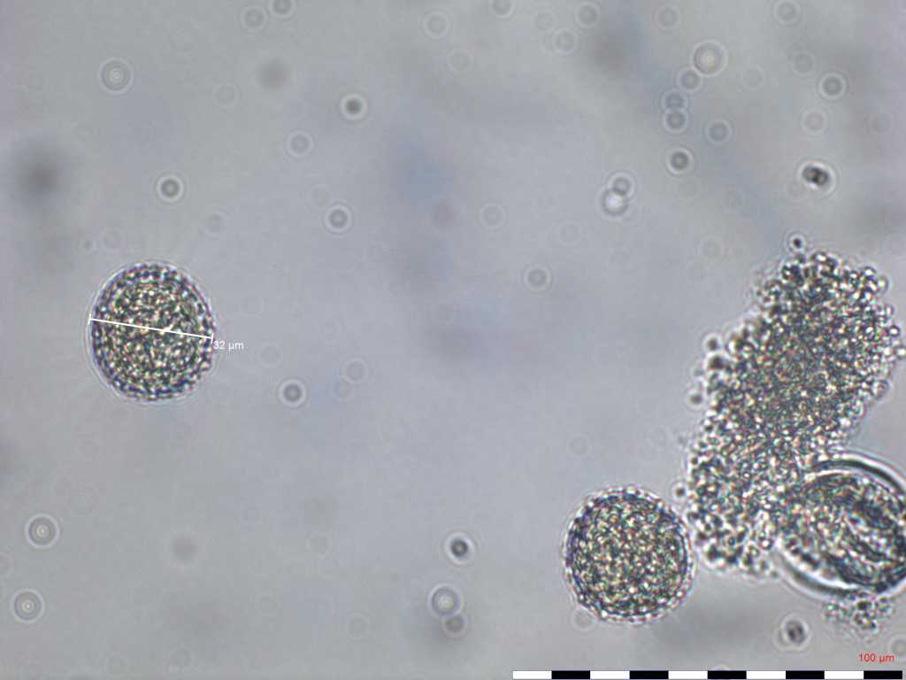
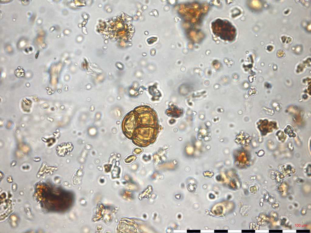
Pollen analysis
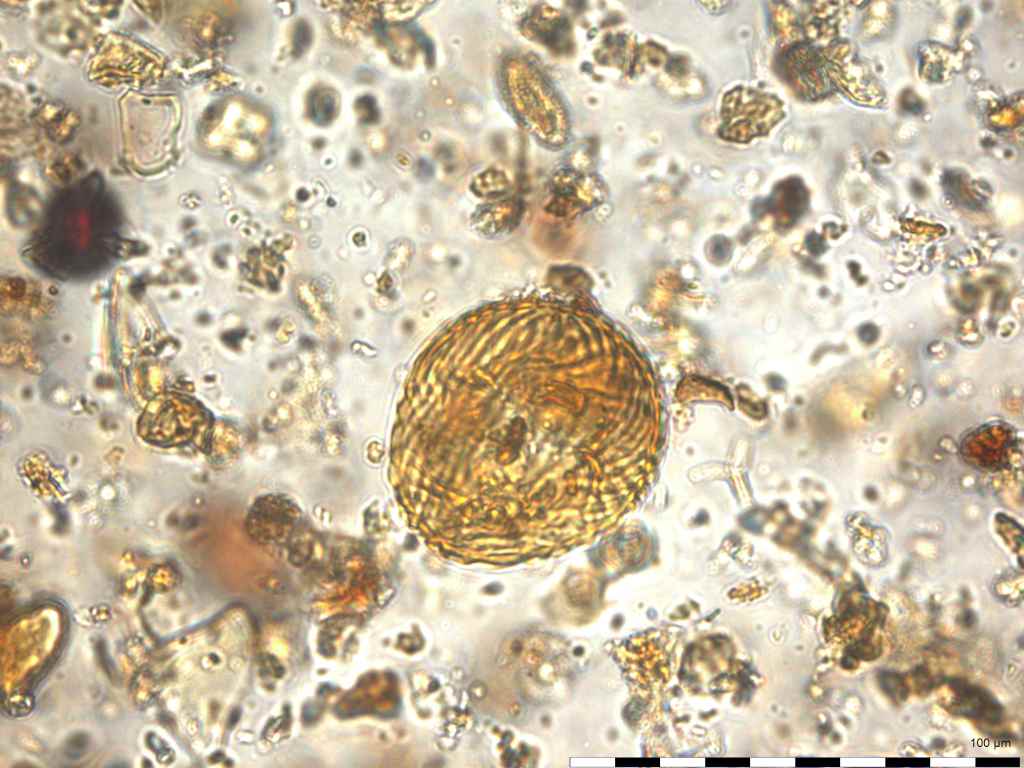
Pollen analysis (palynology) is defined as a method which studies qualitatively and quantitatively recent as well as fossil pollen grains and spores.
The analysis of pollen grains and spores of the Lusatian Miocene lignite seams is applied as an additional method to cuticular analysis. Here, facies identification and stratigraphic classification of lignite seams, layers and corollary horizons have priority.
To receive a profound overview of Tertiary pollen and spores and to preserve a method practiced over many decades, initial training in early Tertiary pollen stratigraphy takes place at the moment. The biostratigraphic evaluation follows the publication of W. Krutzsch concerning the stratigraphic usable spores and pollen forms of the early Tertiary.
The preparation of the samples is realized in our laboratory in Lauta. We are specialized in lignite and carbon-rich samples. The preparation takes place by the use of various chemicals to macerate organic components. For obtaining a maximal enrichment of pollen grains and spores, we endeavor to integrate the preparation of the sample material with fluoric acid into the process. After the chemical preparation the production of permanent preparation with glycerin gelatin follows. Later on these are analyzed by microscope using a magnification of 400 to 600x.
Applications of pollen analysis
- Facies identification of Tertiary lignite
- Biostratigraphy: stratigraphic classification of lignite seams, layers and corollary horizons
- Representation and investigation of fine structures with the aid of the ScanningElectronMicroscope
- Answer to specific questions like:
- Classification of a sample as Tertiary or Quarternary
- Identification of pollen grains in natural honey
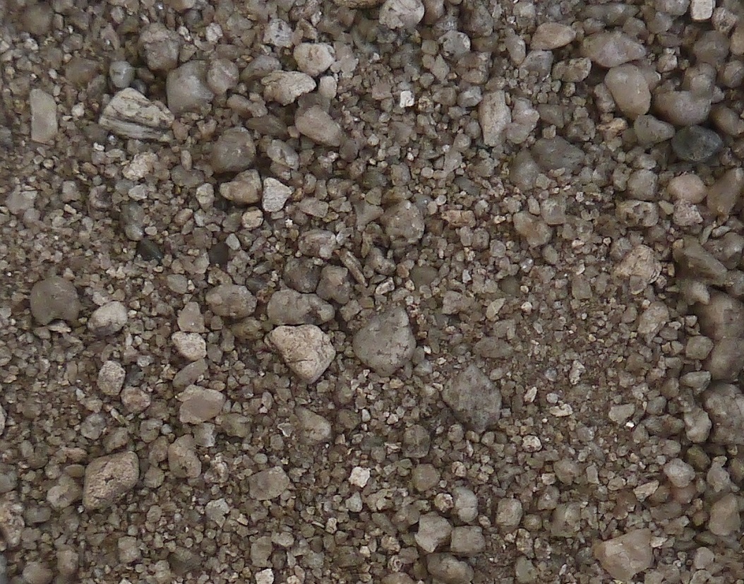
Sediment petrography
Additional to the classic heavy mineral analysis, by using transmitted-light microscopy, the identification of heavy minerals can be extended for opaque minerals by using scanning electron microscopy. With it, different geochemical analyses are possible as well, like for differentiation of various garnet types or for determination of trapped trace elements. In scattered preparation the surface of minerals can be observed. At this, shape, roundness, crack formation, phenomena of dissolution and aggregation of grains can be documented. Such investigation (geochemistry and surface) takes place for light minerals, too. Further, these are described by thin section analysis, where the main focus is on the different quartz formations and feldspar varieties.
The mobile XRF is used for spotty measurement, measurement of samples or generation of series of measurements at drill cores. In the latter case, for instance, an element screening for geochemical characterization of sedimentological sequences can be performed.
For samples with the composition of coarse sand up to gravel, a description will be done using stereomicroscopy. At that, composition, roundness, surface texture and color can be determined. Further a counting of the pebble content in line with TGL 25232 is possible.
Independently of the grain size, each sample can be put through particle analysis with digital image evaluation.
Applications of Sediment Petrography
- Petrographic characterization of sediments
- Make a point about stratigraphy, which can be indicated by mineral changes within a drill core or geological section
- Investigation of the paleogeographic development of sedimentation areas, including conclusions on delivery area, transport, tectonics, diagenesis and climate
- Micro chemical classification of the composition of heavy minerals as an aid for provenance analysis or for the prospection of placer deposits
- Determination of grain size independent enrichment or depletion of, for instance, heavy minerals like zircon
- Determination of the content of rare earth elements, which are preferably enriched in heavy mineral placer
Examples for sediment petrographic researches are given here.